Muscles Labeled Front And Back / Kids Health Topics Your Muscles / Within this group of back muscles you will find the latissimus dorsi, the trapezius, levator scapulae and the rhomboids.
Ditulis oleh
Dorance
Selasa, 09 Februari 2021
Edit
Muscles Labeled Front And Back / Kids Health Topics Your Muscles / Within this group of back muscles you will find the latissimus dorsi, the trapezius, levator scapulae and the rhomboids.. The external intercostal muscles, or external intercostals (intercostales externi) are eleven in number on both sides. Labeled educational inner organ structure. The muscles of the anterior of the forearm are generally divided into two groups: Commonly, people's front delt is significantly more developed than the back portion. For instance the quadriceps muscle group will extend the knee and flex the hip.
Learn the muscles of the leg fast with these quizzes, diagrams and labeling exercises : Broadly considered, human muscle—like the muscles of all vertebrates—is often divided into striated muscle. It is responsible for extension,adduction, and (medial) internal rotation of the shoulder joint. Leg muscle anatomical structure, labeled front, side and back view diagrams. Labeled tick bite infection symptoms scheme.
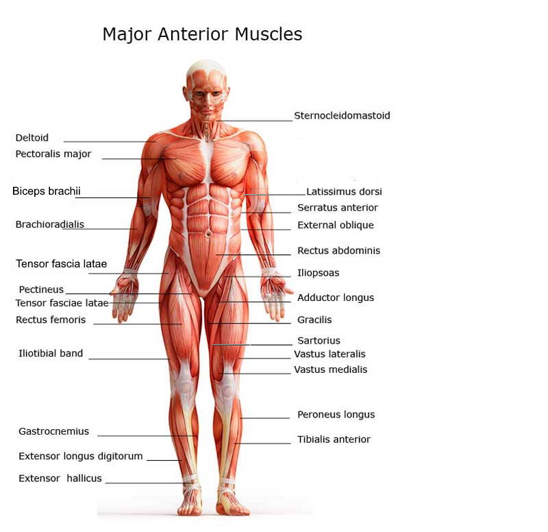
Chart Of Major Muscles On The Front Of The Body With Labels from www.healthpages.org Rotator cuff muscle with anatomical posterior and anterior view expample. Labeled anatomical ear structure scheme. Aalso known as the six pack, is a paired muscle running vertically on each side of the front wall of the abdomen. This labeled human muscular system chart illustrates the major muscle groups in the back (posterior) view and the front (anterior) view. The anterior muscles of the torso (trunk) are those on the front of the body, including the muscles of the chest, abdomen, and pelvis. Click on the labels below to find out more about your muscles. Virus disease symptoms and spreads infographic. Both of these exercises will engage the back portion of your deltoid.
12 photos of the muscles labeled front and back.
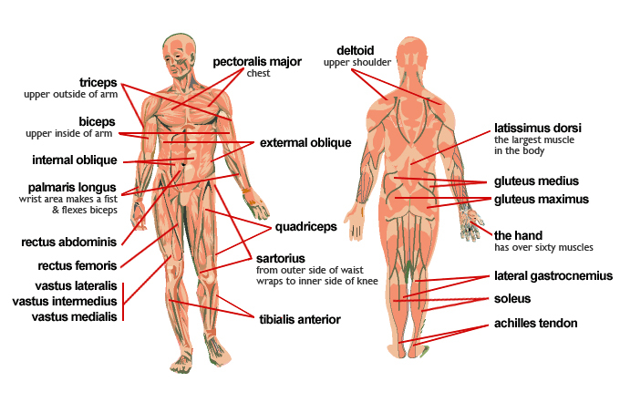
Label the following anatomicalsites in the diagram: Skeletal muscle groups front and back. Your deltoid muscle at your shoulder has a front, middle, and rear part to it. Muscles vary greatly in their shape and size. Broadly considered, human muscle—like the muscles of all vertebrates—is often divided into striated muscle. Male muscular system, full anatomical body diagram with muscle scheme, vector illustration educational poster. The biggest muscle is lats muscle, then there are traps muscle. Commonly, people's front delt is significantly more developed than the back portion. Triceps, biceps, pectoralis major, quadriceps , hamstrings, gluteus maximus , abdominals, deltoid, latissimus dorsi, external obliques, gastrocnemius , tibialis anterior. The trapezius originates from the skull and spine of the. Attachments, nerve supply well there are lot of muscles on back and every muscle is trained differently. This labeled human muscular system chart illustrates the major muscle groups in the back (posterior) view and the front (anterior) view. Want to learn more about it?
Back view of muscles, skeleton, organs, nervous system. Virus disease symptoms and spreads infographic. Front view of woman's thigh and knee muscles with names. Attachments, nerve supply well there are lot of muscles on back and every muscle is trained differently. The muscles extend from the tubercles of the ribs behind, to the cartilages of the ribs in front, where they end in thin membranes, the external intercostal membranes.
Pin On The Class from i.pinimg.com A back muscle that pulls the arm down and back. Tutorials and quizzes on the anatomy and actions of the back muscles (iliocostalis, longissimus, spinalis, multifidus, and quadratus lumborum), using interactive animations, diagrams, and illustrations. This muscular system chart shows in detail the deep layers of muscle on the back side of your body. Skeletal muscle groups front and back. The muscle fibers' highly specialized structure enables the muscles to relax and contract to produce movement. Click on the labels below to find out more about your muscles. Commonly, people's front delt is significantly more developed than the back portion. Front view of woman's thigh and knee muscles with names.
Skeletal muscle groups front and back.
Attachments, nerve supply well there are lot of muscles on back and every muscle is trained differently. Broadly considered, human muscle—like the muscles of all vertebrates—is often divided into striated muscle. Vector illustration informative medical scheme. Human muscle system, the muscles of the human body that work the skeletal system, that are under voluntary control, and that are concerned with movement, posture, and balance. The muscles extend from the tubercles of the ribs behind, to the cartilages of the ribs in front, where they end in thin membranes, the external intercostal membranes. Label the following anatomicalsites in the diagram: Virus disease symptoms and spreads infographic. A back muscle that pulls the arm down and back. Labeled anatomical ear structure scheme. Your deltoid muscle at your shoulder has a front, middle, and rear part to it. The superficial back muscles are the muscles found just under the skin. The anterior muscles of the torso (trunk) are those on the front of the body, including the muscles of the chest, abdomen, and pelvis. Labeled educational inner organ structure.
Back of the head muscle structure and nerve system diagram. Labeled educational inner organ structure. This labeled human muscular system chart illustrates the major muscle groups in the back (posterior) view and the front (anterior) view. Tutorials and quizzes on the anatomy and actions of the back muscles (iliocostalis, longissimus, spinalis, multifidus, and quadratus lumborum), using interactive animations, diagrams, and illustrations. Back view of muscles, skeleton, organs, nervous system.
Meet Some Muscles Science Learning Hub from static.sciencelearn.org.nz Tutorials and quizzes on the anatomy and actions of the back muscles (iliocostalis, longissimus, spinalis, multifidus, and quadratus lumborum), using interactive animations, diagrams, and illustrations. Virus disease symptoms and spreads infographic. Related posts of muscles labeled front and back. Text and images from slide. Want to learn more about it? Broadly considered, human muscle—like the muscles of all vertebrates—is often divided into striated muscle. Commonly, people's front delt is significantly more developed than the back portion. The muscle fibers' highly specialized structure enables the muscles to relax and contract to produce movement.
Both of these exercises will engage the back portion of your deltoid.
Labeled anatomical ear structure scheme. Both of these exercises will engage the back portion of your deltoid. Text and images from slide. 12 photos of the muscles labeled front and back. It is responsible for extension,adduction, and (medial) internal rotation of the shoulder joint. Click on the labels below to find out more about your muscles. We have more than 600 individual muscles in our body, and although you're not responsible for knowing the muscle anatomy in this class, knowing that anatomy and knowing the different muscle groups and how they work. This muscular system chart shows in detail the deep layers of muscle on the back side of your body. Each of your muscles is made up of thousands of thin, long, cylindrical cells called muscle fibers. Commonly, people's front delt is significantly more developed than the back portion. Skeletal muscle groups front and back. Labeled educational inner organ structure. Liver inflammation with scar tissues and cirrhosis.
Ihsandroid
Jika Bermanfaat Mohon Donasi Se-Iklasnya via: OVO (0812 1048 8454) / DANA (0812 1048 8454) Kontak Saya Via Whatsapp, Terima kasih kepada anda yang telah berkunjung ke Website ini dan memberikan Donasi.